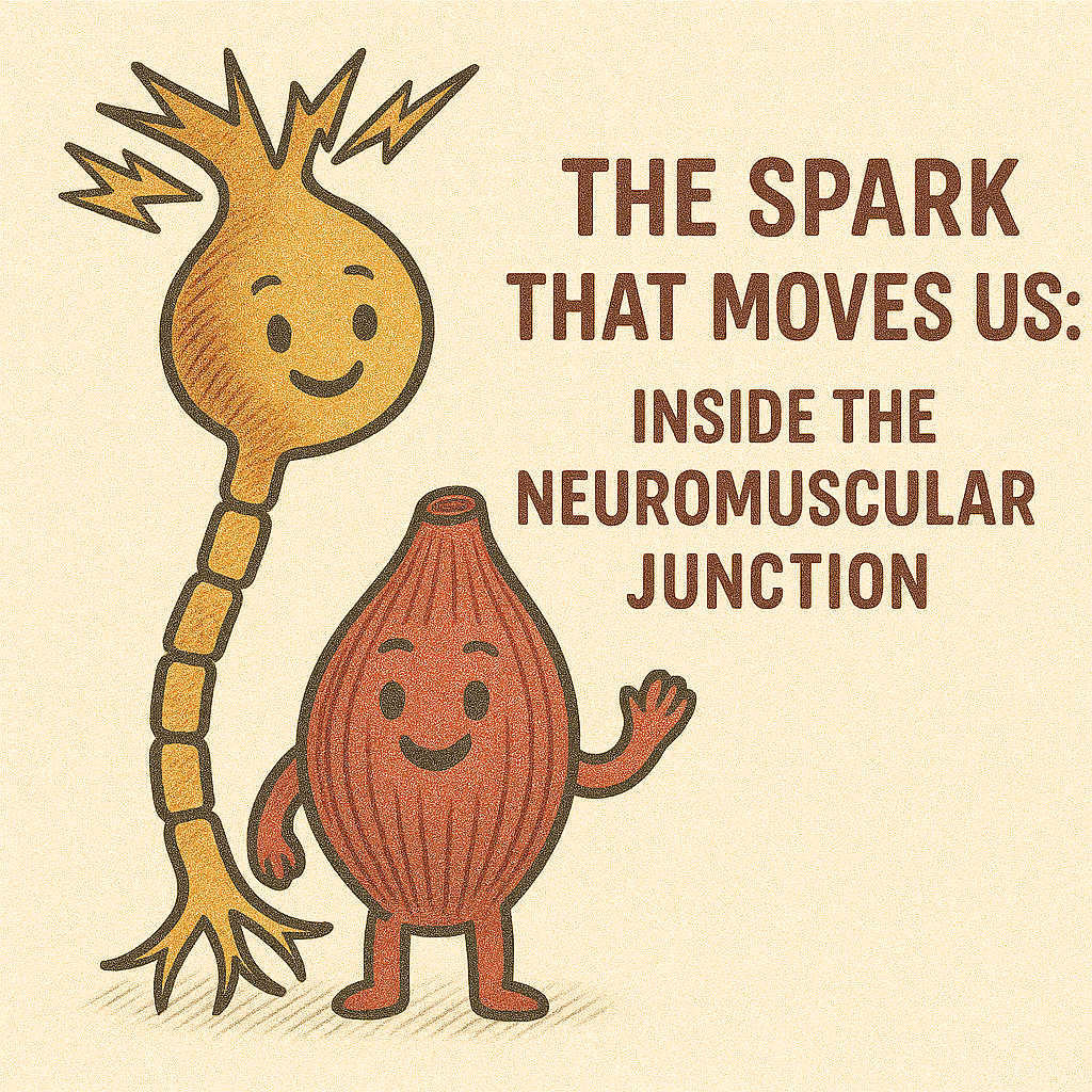
Where electricity becomes motion, and thought turns into action.
🌟 Prelude — The Moment Before Movement
Before your hand ever touches a scalpel or your foot presses the accelerator, a silent messenger races through your nerves. In that blink of time, neuromuscular transmission converts a neuron’s whisper into a muscle’s roar.
This invisible dialogue between nerve and muscle lies at the heart of motion, anesthesia, and even life itself. Understanding it is like learning the grammar of movement — precise, elegant, and endlessly fascinating.
🧩 The Connection — Anatomy of the Neuromuscular Junction (NMJ)
Picture a high-security handshake 🤝 between two elite partners:
- The presynaptic terminal of a motor neuron, brimming with neurotransmitter-filled vesicles.
- The synaptic cleft, a microscopic gap (~50 nm) bridging the two.
- The postsynaptic membrane, or motor endplate, studded with nicotinic acetylcholine receptors (nAChRs).
Surrounding this zone are Schwann cells (insulating the signal) and a basal lamina rich in acetylcholinesterase — the molecule that will later silence the conversation.
🧠 Concept Box
Each motor neuron may command hundreds of muscle fibres, yet every muscle fibre obeys only one neuron — ensuring loyalty in motion.
⚙️ The Sequence — How the Signal Travels
When you decide to move, a cascade begins:
- ⚡ Action potential races down the motor neuron.
- 🧲 Voltage-gated Ca²⁺ channels open at the nerve terminal.
- 💬 Acetylcholine (ACh) is released into the synaptic cleft.
- 🧩 ACh binds to nicotinic receptors → Na⁺ rushes in → local depolarization (end-plate potential).
- 🎯 If threshold is reached, a muscle action potential fires along the sarcolemma and into T-tubules, triggering Ca²⁺ release from the sarcoplasmic reticulum.
- 💪 Cross-bridge cycling begins — and movement is born.
💊 Clinical Pearl
Non-depolarizing relaxants (e.g., vecuronium, rocuronium) block ACh receptors.
Depolarizing agents (e.g., succinylcholine) mimic ACh but resist breakdown, keeping the gate open until paralysis sets in.
🕊️ Termination — The Art of Silence
Every conversation must end, even between nerve and muscle.
Once contraction begins, acetylcholinesterase (AChE) rapidly hydrolyses ACh into acetate and choline within milliseconds. Choline is then recycled into the neuron for another round.
⚠️ Caution Box
When AChE is inhibited — as in organophosphate poisoning — ACh accumulates, causing continuous depolarization and potentially lethal paralysis.
💊 Clinical Pearl (Green)
Neostigmine (a reversible AChE inhibitor) extends ACh activity, helping reverse non-depolarizing neuromuscular blockade at the end of surgery.
🧱 Reliability in Motion — Safety Factor and Synaptic Reserve
The NMJ is built with redundancy. Even if a few vesicles fail to release, enough ACh normally floods the cleft to ensure depolarization — this is the safety factor.
Behind this reliability is a reserve pool of vesicles and the principle of quantal release, where each vesicle’s content forms one “quantum” of transmitter effect.
🧠 Concept Box
At rest, mini end-plate potentials (MEPPs) flicker like tiny sparks — evidence of spontaneous vesicle fusion, keeping the system “primed.”
📚 Exam Corner
↓ Safety factor → fatigable weakness (seen in myasthenia gravis).
↑ Quantal content → enhanced transmission (seen after cholinesterase inhibitors).
⚠️ When the Spark Falters — Disorders of Neuromuscular Transmission
| ⚕️ Disorder | 🧬 Mechanism | 🧠 Key Clinical Features |
|---|---|---|
| Myasthenia Gravis | Autoantibodies destroy postsynaptic ACh receptors | Fatigable weakness, ptosis, improved by rest |
| Lambert–Eaton Syndrome | Antibodies block presynaptic Ca²⁺ channels | Proximal weakness, reduced ACh release |
| Botulism | Toxin cleaves SNARE proteins → ACh release blocked | Flaccid paralysis, respiratory failure |
| Organophosphate Poisoning | AChE inhibition → ACh excess | Miosis, bradycardia, fasciculations, paralysis |
💊 Clinical Pearl
Botulinum toxin, despite its fearsome nature, is therapeutically harnessed in microdoses for dystonias and cosmetic relaxation.
⚠️ Caution Box
In anesthetic practice, always differentiate myasthenic vs cholinergic crisis — both cause weakness but demand opposite treatments.
😴 Under the Anesthesiologist’s Lens — NMJ and Muscle Relaxants
In the operating theatre, the NMJ becomes the anesthesiologist’s stage. Muscle relaxants let surgery proceed with precision, but their management demands respect for physiology.
💊 Clinical Pearl
- Depolarizing agents (e.g., succinylcholine): mimic ACh → sustained depolarization → transient fasciculations → paralysis.
- Non-depolarizing agents (e.g., atracurium, rocuronium): competitively block ACh binding → paralysis without fasciculations.
- Reversal: Neostigmine + Glycopyrrolate or Sugammadex (for aminosteroids).
🧠 Concept Box
Train-of-Four (TOF) monitoring measures fade in twitch amplitude to assess residual paralysis — a TOF ratio ≥ 0.9 signals safe recovery.
📚 Exam Corner
PORC (Postoperative Residual Curarization) remains a significant risk if reversal is incomplete.
🚀 Horizons Ahead — Neuromuscular Research in Motion
Modern physiology peers deeper than ever:
- 🔬 Super-resolution imaging reveals single ACh vesicle fusion in real time.
- 🧬 Genomic studies uncover mutations in AChR subunits causing congenital myasthenic syndromes.
- 💉 Biologics such as eculizumab and ravulizumab offer new hope in refractory MG.
- 👩🚀 Space medicine explores NMJ adaptation in microgravity, redefining human performance beyond Earth.
💊 Clinical Pearl
Future neuromuscular therapies may involve synaptic gene editing and bioengineered receptor regeneration — restoring lost dialogue between nerve and muscle.
🌌 The Whisper Between Nerve and Muscle
At every instant, trillions of these microscopic sparks maintain our breath, blink, and balance.
The neuromuscular junction is not just a synapse — it’s a negotiation of will, the physical manifestation of intent. From the surgeon’s steady hand to a newborn’s first cry, it reminds us that communication defines life.
✨ “Between thought and action lies a whisper — the whisper of electricity meeting flesh.”
External References: PubMed, StatPearls, Frontiers in Physiology
Leave a Reply