“Every alveolus is a stage, every red cell a dancer — and the rhythm of life depends on how perfectly they meet.”
🌬️ The Hidden Choreography of Breath
Take a deep breath — feel the cool air swirl down your trachea, branching through bronchi like rivers through valleys, until it reaches millions of tiny air sacs.
Here, in the quiet intimacy of an alveolus, air and blood perform a silent ballet. Oxygen flows in, carbon dioxide drifts out, and life continues — unannounced but unwavering.
Yet, this exchange is only as graceful as the balance between two essential partners:
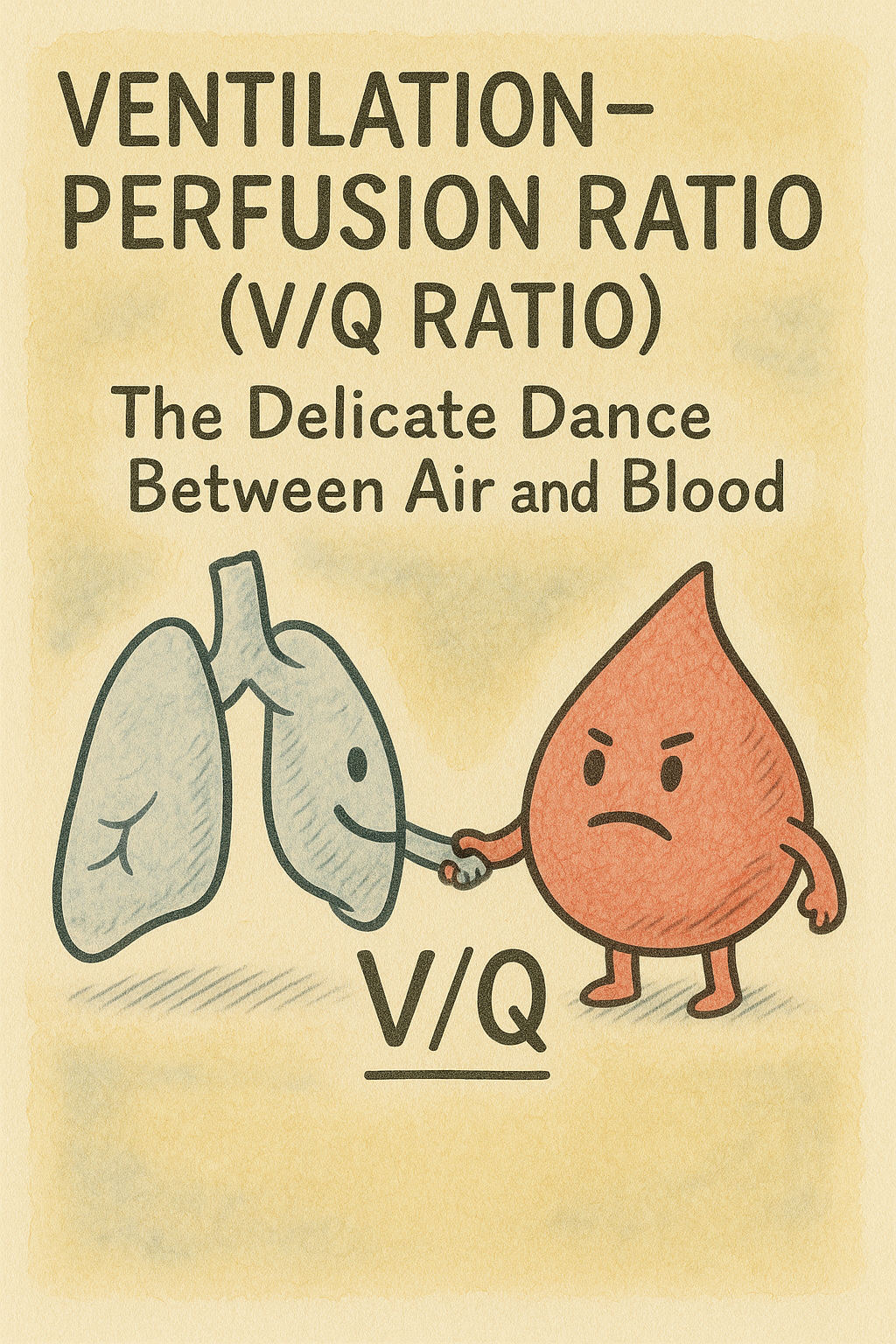
- Ventilation (V): the air that reaches the alveoli.
- Perfusion (Q): the blood that flows through pulmonary capillaries.
Together, they define the ventilation–perfusion ratio, or V/Q ratio — a concept that decides whether a breath truly serves its purpose.
The Equation of Balance
Physiology often hides poetry in equations. The V/Q ratio is one such equation:
V/Q = Ventilation (L/min) / Perfusion (L/min)
Under normal resting conditions:
- Alveolar ventilation ≈ 4 L/min
- Pulmonary blood flow ≈ 5 L/min
Hence, the normal V/Q ratio ≈ 0.8 — a delicate equilibrium that sustains life.
But this balance is far from uniform. Gravity, posture, and local pressures ensure that some parts of the lung are better ventilated or perfused than others. And that’s where the story becomes fascinating.
(💡 Metaphor: “The lungs are like a mountain — the air is plenty at the summit, the blood richer at the base.”)
🌍 Gravity’s Influence — The Lung as an Inverted Tree
Imagine the lung as an inverted tree, roots up, branches down.
At the base, gravity pulls both air and blood downward, but blood — being heavier — flows more.
At the apex, ventilation still occurs, but perfusion lags behind.
The result:
- Apex: High V/Q ratio (ventilation > perfusion) → alveoli are well-aerated but underperfused → wasted ventilation or physiological dead space.
- Base: Low V/Q ratio (perfusion > ventilation) → alveoli are well-perfused but poorly ventilated → shunt-like effect.
This uneven distribution means that while oxygen diffuses efficiently at the base, the apex contributes more to carbon dioxide elimination.
(🌿 Poetic insight: “The lungs are democratic — yet gravity gives each region a different vote.”)
⚖️ The Zones of the Lung — West’s Model
Physiologist John West elegantly described the lung as divided into zones, based on the relationships among alveolar (PA), arterial (Pa), and venous (Pv) pressures.
Let’s travel through them like explorers moving down a mountain:
- Zone I (PA > Pa > Pv):
- Alveolar pressure compresses capillaries.
- No perfusion occurs — alveolar dead space.
- Seen in hypovolemia or high positive pressure ventilation.
- Zone II (Pa > PA > Pv):
- Blood flow is intermittent, like a waterfall.
- Flow depends on the difference between Pa and PA.
- Zone III (Pa > Pv > PA):
- Continuous perfusion — ideal gas exchange.
- This is where most of oxygenation occurs in healthy lungs.
- Zone IV (pathological):
- Occurs in disease states with reduced compliance and edema.
These zones remind us that the lung is not a static organ — it’s a dynamic terrain where physics dictates physiology.
💨 When the Dance Falters — V/Q Mismatch
The V/Q mismatch occurs when ventilation and perfusion are not proportionally matched. This is the most common cause of hypoxemia in both anesthesia and critical care.
Let’s dissect it:
- Low V/Q ratio:
- Alveoli are perfused but poorly ventilated.
- Common in conditions like pneumonia, asthma, or atelectasis.
- Leads to hypoxemia that improves with oxygen therapy.
- High V/Q ratio:
- Alveoli are ventilated but poorly perfused.
- Seen in pulmonary embolism or emphysema.
- Represents wasted ventilation.
- V/Q = 0:
- No ventilation, only perfusion → shunt.
- Seen in ARDS or complete airway obstruction.
- V/Q = ∞:
- No perfusion, only ventilation → dead space.
- Seen in pulmonary embolism.
(💫 “When air arrives to find no blood, or blood comes to find no air — the harmony fades.”)
🩺 Clinical Illustrations — When Physiology Meets Reality
Case 1: The Silent Base
A 45-year-old man post-abdominal surgery lies supine, his shallow breathing promoting atelectasis at lung bases.
His oxygen saturation drops, and his arterial blood gas shows low PaO₂ but normal PaCO₂ — a classic low V/Q scenario.
Re-expanding his alveoli with physiotherapy and low-level PEEP restores oxygenation.
Case 2: The Lost Perfusion
A trauma patient develops a pulmonary embolism.
His alveoli are open, but blood flow is obstructed — a high V/Q mismatch.
Despite high oxygen, PaO₂ remains low until the clot is lysed.
🧪 The Alveolar–Arterial Gradient — Measuring the Inefficiency
The A–a gradient quantifies how well oxygen moves from alveoli to arteries.
A–a Gradient = PAO₂ – PaO₂
The alveolar gas equation helps calculate PAO₂:
PAO₂ = FiO₂ × (Pb – PH₂O) – (PaCO₂ / R)
- Normal A–a gradient: 5–15 mmHg (young) to 20–30 mmHg (elderly).
- Increased A–a gradient: seen in V/Q mismatch, diffusion defects, or shunt.
Example:
In pneumonia (low V/Q), PAO₂ may remain high, but PaO₂ drops → widened A–a gap → impaired oxygen diffusion efficiency.
⚙️ V/Q and Mechanical Ventilation — The Art of Balance
Mechanical ventilation is like conducting an orchestra — every parameter affects the symphony of gas exchange.
- Positive End-Expiratory Pressure (PEEP):
- Recruits collapsed alveoli → improves low V/Q.
- Excessive PEEP, however, overdistends alveoli → increases high V/Q.
- Tidal Volume:
- Too low → alveolar collapse.
- Too high → volutrauma and V/Q imbalance.
- Positioning:
- Prone ventilation in ARDS redistributes perfusion, improving oxygenation.
(⚙️ Clinical pearl: In anesthesia, muscle relaxation and supine posture reduce FRC and predispose to low V/Q mismatch — hence the need for optimal PEEP and recruitment maneuvers.)
V/Q Scanning and Imaging
In nuclear medicine, the V/Q scan is the diagnostic equivalent of watching the lungs dance on screen.
- Ventilation scan: patient inhales radioactive gas (e.g., Xenon or Tc-99m).
- Perfusion scan: intravenous radiotracer maps pulmonary blood flow.
- Mismatch areas: highlight pulmonary embolism — ventilated regions with poor perfusion.
Modern imaging like dual-energy CT and electrical impedance tomography now allows real-time visualization of V/Q distribution in ventilated patients.
🌫️ Hypoxic Pulmonary Vasoconstriction — The Lung’s Self-Correction
Even when the V/Q ratio wavers, the lungs have a built-in guardian — hypoxic pulmonary vasoconstriction (HPV).
When alveolar oxygen tension drops, local arterioles constrict, diverting blood toward better-ventilated areas.
This reflex preserves overall V/Q matching and limits shunt fraction.
However, HPV can be inhibited by:
- Volatile anesthetics
- Sepsis
- Alkalosis
- Excessive vasodilators
(💭 “Even in imbalance, the lungs seek harmony — tightening their vessels like strings on an instrument until the rhythm returns.”)
🌍 At the Extremes — High Altitude and Diving
High Altitude:
At altitude, hypoxia triggers global HPV, raising pulmonary vascular resistance → possible pulmonary hypertension.
Yet, this adaptive response helps redirect blood flow for improved oxygen uptake in limited air pressure.
Diving:
In deep-sea diving, increased ambient pressure raises gas density → alters airflow resistance → affects regional ventilation.
Here, perfusion dominates and V/Q ratio falls, risking carbon dioxide retention.
“The mountain’s summit and the ocean’s floor tell the same story — the fragile balance between air and blood.”
🧠 Integrative Summary Table
| Condition | V/Q Ratio | Effect | PaO₂ | PaCO₂ | Response to O₂ |
|---|---|---|---|---|---|
| Normal | 0.8 | Optimal exchange | Normal | Normal | N/A |
| Pulmonary Embolism | ↑ | Dead space | ↓ | ↓ | Improves |
| Pneumonia | ↓ | Shunt-like | ↓↓ | ↑ | Partial |
| Emphysema | Variable ↑ | Wasted ventilation | ↓ | ↓ | Partial |
| ARDS | ↓ | Severe mismatch | ↓↓ | ↑ | Partial |
| Atelectasis | ≈ 0 | True shunt | ↓↓ | ↑ | Poor |
🌟 Key Takeaways
✅ The V/Q ratio defines the balance between air flow and blood flow.
✅ Normal ≈ 0.8, but varies regionally due to gravity and perfusion gradients.
✅ Low V/Q → shunt-like, High V/Q → dead-space-like patterns.
✅ A–a gradient quantifies oxygenation inefficiency.
✅ HPV helps restore V/Q balance but is impaired in anesthesia.
✅ Clinical scenarios: ARDS, PE, COPD, atelectasis, pneumonia.
✅ Ventilator strategy = the art of keeping air and blood in perfect conversation.
🌸 Closing Reflection
“In the lungs, perfection is not uniformity — it is balance.
When air finds blood in just the right measure, life sings its softest song.”
References:
- Guyton & Hall. Textbook of Medical Physiology, 14th Edition.
- West JB. Respiratory Physiology: The Essentials, 11th Edition.
- Nunn’s Applied Respiratory Physiology, 9th Edition.
- Miller’s Anesthesia, 9th Edition.
- American Journal of Respiratory and Critical Care Medicine.
- Chest Journal, Critical Care Medicine.
✨ “The balance between air and blood is not just physiology — it’s poetry written in pressure and flow.” ✨
Leave a Reply