“Between every breath lies a border — air on one side, blood on the other — and in that silent crossing, life renews itself.”
🌬️ The Border Where Atmosphere Meets Life
Take a slow breath. The air you just drew in carries the same oxygen that once touched mountain peaks and ocean waves. Within seconds, it will reach the alveolar–capillary membrane, an invisible interface only a few micrometers thick, yet powerful enough to sustain existence.
At this border, the diffusion of gases occurs — a masterpiece of natural engineering that turns every breath into chemistry and every heartbeat into continuity.
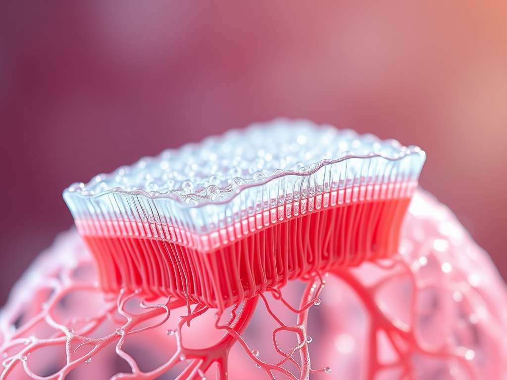
🧬 The Architecture of an Invisible Wall
The alveolar–capillary membrane is a multilayered interface designed for perfection. Despite its fragility, it offers a vast area — nearly 70 square metres, roughly the size of a tennis court. Across this surface, more than 300 million alveoli open like microscopic balloons, welcoming air to dance with blood.
Let us walk through its layers — the path a molecule of oxygen must cross:
- A thin surfactant film that lowers surface tension.
- The alveolar epithelium (type I and II pneumocytes).
- The epithelial basement membrane.
- A narrow interstitial space containing fibroblasts and collagen.
- The capillary basement membrane.
- Finally, the capillary endothelium, before diffusing into plasma and erythrocytes.
All of this is barely 0.3 micrometres thick — thinner than a spider’s silk. Yet through this delicate veil, every molecule of oxygen enters the bloodstream.
(💭 “The thinnest wall in the body guards the greatest secret of life.”)
⚖️ Fick’s Law — The Mathematics of Breath
Nature obeys laws, even in poetry. The movement of gases across this membrane follows Fick’s Law of Diffusion:
Rate of Diffusion = (A × D × ΔP) / T
Where:
- A = surface area
- D = diffusion coefficient (related to solubility / molecular weight)
- ΔP = partial pressure difference across the membrane
- T = thickness of the barrier
Oxygen and carbon dioxide differ not by intention but by physics:
- Carbon dioxide is about 20 times more soluble than oxygen.
- Oxygen, however, has a higher partial-pressure gradient, allowing rapid entry despite lower solubility.
Thus, while carbon dioxide slips through easily, oxygen’s speed is driven by a stronger push.
🌬️ Partial Pressures — The Engines of Movement
Gas molecules move not because they want to, but because pressure tells them to.
| Location | PO₂ (mm Hg) | PCO₂ (mm Hg) |
|---|---|---|
| Alveolar air | 104 | 40 |
| Pulmonary capillary (venous) | 40 | 45 |
| Pulmonary capillary (arterial end) | 95 | 40 |
Within only 0.25 seconds — half the time blood spends in the capillary — oxygen equilibrates with alveolar air. Carbon dioxide, despite its lower gradient, completes its journey even faster thanks to its higher solubility.
(🌿 Metaphor: “It’s a quiet conversation — oxygen whispering in, carbon dioxide sighing out.”)
🔬 Diffusion-Limited vs Perfusion-Limited Exchange
Not all gases cross with the same limitation. Two fundamental scenarios define their journey:
Perfusion-limited diffusion
- The gas equilibrates quickly; further uptake depends on blood flow.
- Example: Nitrous oxide (N₂O) — as soon as it enters blood, pressures equalise.
Diffusion-limited diffusion
- The gas never reaches equilibrium; transfer depends on membrane properties.
- Example: Carbon monoxide (CO) — it binds instantly to haemoglobin, keeping blood partial pressure low.
Oxygen is typically perfusion-limited but can become diffusion-limited in diseases like fibrosis, edema, or during high-altitude hypoxia.
🧪 Diffusing Capacity of the Lung — Measuring the Magic
Clinically, we quantify this phenomenon as the Diffusing Capacity of the Lung for Carbon Monoxide (DLCO):
DLCO = Rate of CO transfer / (Alveolar–capillary pressure difference)
Normal DLCO ≈ 25 mL/min/mm Hg at rest.
| Condition | DLCO Change | Reason |
|---|---|---|
| Emphysema | ↓ | Loss of alveolar surface area |
| Pulmonary fibrosis | ↓ | Thickened membrane |
| Pulmonary edema | ↓ | Fluid barrier |
| Polycythemia | ↑ | More haemoglobin binding sites |
| Exercise | ↑ | Increased capillary recruitment |
Because carbon monoxide’s diffusion is membrane-limited, its uptake reflects the integrity and efficiency of the alveolar–capillary barrier.
🩸 Diseases That Distort the Diffusion Ballet
When the lung’s choreography falters, diffusion suffers — and hypoxemia follows.
Pulmonary Fibrosis
Collagen thickens the interstitium, increasing diffusion distance. Patients desaturate early during exertion because oxygen cannot equilibrate in shortened transit time.
Emphysema
Alveolar destruction reduces surface area. Oxygen diffusion falls despite normal membrane thickness — an architectural failure.
Pulmonary Edema
Fluid floods the interstitial and alveolar space, diluting gradients and thickening barriers.
Anemia
Diffusion across the membrane remains intact, but oxygen-carrying capacity drops; the blood becomes an empty courier.
🧍♂️ The Diffusion Reserve — Nature’s Safety Margin
At rest, a red blood cell spends 0.75 seconds in a pulmonary capillary, but equilibrium occurs in about 0.25 seconds.
This spare time forms the diffusion reserve — a safety buffer allowing gas exchange even during exercise or mild disease.
During maximal exertion, capillary transit may shorten to 0.25 seconds. Healthy lungs manage; diseased lungs do not — leading to exercise-induced desaturation.
(💫 “Even in haste, the lungs make time for exchange — until disease steals the seconds.”)
⚙️ Clinical Relevance — When Physiology Meets Practice
Understanding diffusion guides our clinical decisions daily.
- Oxygen therapy: Increases the partial-pressure gradient (ΔP), enhancing oxygen uptake.
- Positive end-expiratory pressure (PEEP): Recruits collapsed alveoli, restoring surface area.
- High altitude: Reduced barometric pressure lowers ΔP; hence supplemental oxygen or acclimatisation becomes essential.
- Anesthesia: Low functional residual capacity and micro-atelectasis decrease effective surface area for diffusion.
(🩺 Clinical Tip: Patients with diffusion defects respond to high FiO₂ if the defect is purely membrane-based, but not if perfusion is also compromised.)
🌍 Diffusion at the Extremes
High Altitude — The Thin Air Challenge
At 5 000 metres, barometric pressure drops to nearly half of sea-level values. Even with perfect lungs, ΔP for oxygen plummets, slowing diffusion. The body compensates with hyperventilation and erythropoietin-driven polycythemia.
Exercise — The Accelerated Exchange
In elite athletes, cardiac output may rise five-fold, cutting transit time drastically. Healthy membranes cope; diseased ones reveal their weakness.
Anesthesia — The Controlled Compromise
Supine position and muscle relaxation reduce functional residual capacity, promoting small-airway closure and reducing surface area. Diffusion becomes partially compromised unless recruitment manoeuvres are applied.
🧠 Integrative Table — Factors Influencing Gas Diffusion
| Variable | Change | Effect on Diffusion | Example |
|---|---|---|---|
| Surface area (A) ↓ | ↓ Rate | Emphysema | |
| Thickness (T) ↑ | ↓ Rate | Fibrosis, edema | |
| ΔP (gradient) ↓ | ↓ Rate | High altitude | |
| Diffusion coefficient (D) ↓ | ↓ Rate | CO₂ disorders (rare) | |
| Blood volume ↑ | ↑ Rate | Exercise | |
| Haemoglobin ↓ | ↓ O₂ transport | Anemia |
💡 Diffusion vs Perfusion Mismatch — Clinical Intersection
In reality, gas exchange is rarely purely diffusion- or perfusion-limited.
Diseases like COPD or ARDS create regional V/Q differences where both limitations coexist — diffusion impaired, perfusion mismatched.
Understanding this interplay helps optimise mechanical ventilation and oxygen delivery strategies.
(🌾 “When air and blood fall out of rhythm, the song of life turns faint — yet the lungs strive to retune the melody.”)
🌸 Key Takeaways
✅ Gas diffusion in the lungs is governed by Fick’s Law — driven by surface area, membrane thickness, and partial-pressure gradients.
✅ Oxygen diffusion depends on ΔP, while carbon dioxide relies on high solubility.
✅ DLCO quantifies membrane integrity; it falls in emphysema, fibrosis, and edema.
✅ Diffusion reserve prevents hypoxemia during brief stress, but disease erodes this safety net.
✅ Clinical application spans oxygen therapy, PEEP optimisation, and altitude medicine.
🌙 Closing Reflection
“Every heartbeat sends a river; every breath releases wind.
Where they meet, life is forged — unseen, silent, and perfectly balanced.”
📚 References
- West JB. Respiratory Physiology: The Essentials, 11th Edition.
- Guyton & Hall. Textbook of Medical Physiology, 14th Edition.
- Nunn’s Applied Respiratory Physiology, 9th Edition.
- Miller’s Anesthesia, 9th Edition.
- American Thoracic Society. Standards for Single-Breath DLCO Testing, 2022.
- Chest Journal & Critical Care Medicine latest reviews on diffusion capacity and gas exchange.
✨ “Science explains how; poetry reminds us why.” ✨
Leave a Reply